Cai-yun Li,
Zheng-cheng Zhang ![]() ,
Jing-yue Mao,
Li-feng Sho,
Ying Zheng,
Jia-li Quan
,
Jing-yue Mao,
Li-feng Sho,
Ying Zheng,
Jia-li Quan
For correspondence:- Zheng-cheng Zhang Email: zhengceng3476@hotmail.com Tel:+8657187348682
Received: 18 October 2016 Accepted: 14 February 2017 Published: 31 March 2017
Citation: Li C, Zhang Z, Mao J, Sho L, Zheng Y, Quan J. Preparation of Tradescantia pallida-mediated zinc oxide nanoparticles and their activity against cervical cancer cell lines. Trop J Pharm Res 2017; 16(3):494-500 doi: 10.4314/tjpr.v16i3.1
© 2017 The authors.
This is an Open Access article that uses a funding model which does not charge readers or their institutions for access and distributed under the terms of the Creative Commons Attribution License (http://creativecommons.org/licenses/by/4.0) and the Budapest Open Access Initiative (http://www.budapestopenaccessinitiative.org/read), which permit unrestricted use, distribution, and reproduction in any medium, provided the original work is properly credited..
Purpose: To synthesize zinc oxide nanoparticles (ZnO NPs) using Tradescantia pallida. (Commelinaceae) and determine their fluorescent and cytotoxic properties.
Methods: ZnO NPs were synthesized according to a simple protocol using T. pallida aqueous leaf extract (TPALE). Scanning electron microscopy (SEM) and transmission electron microscopy (TEM) were used to analyze the morphology of the ZnO NPs. X-ray diffraction (XRD) and Fourier transform-infrared spectroscopy (FTIR) measurements were performed to determine their crystalline nature and functional groups, respectively. Fluorescence spectroscopy was used to assess the photoluminescence properties of ZnO NPs. Upon confirmation of ZnO NP synthesis, cytotoxicity tests were carried out against HeLa cell line by 3-(4,5-dimethylthiazol-2-yl)-2,5-diphenyltetrazolium bromide (MTT) assay.
Results: The agglomerated ZnO NPs were rod-shaped and had a mean particle size of 25 ± 2 nm. Further, they exhibited good photoluminescence with correlation to ZnO crystals. MTT assay results indicated significant cytotoxicity against HeLa cervical cancer cell line.
Conclusion: A simple approach for ZnO NP synthesis based on TPALE has been developed successfully. The synthesized ZnO NPs demonstrate good luminescence properties and cytotoxicity against cervical cancer line.
Introduction
Nanotechnology is one of the most versatile areas of current research and has wide-ranging applications. A number of toxic physio-chemical techniques, such as spray pyrolysis, gas-phase methods, chemical vapor deposition, electrochemical methods, and laser ablation techniques, have been introduced to synthesize nanoparticles (NPs) [1]. In this study, we focused on non-toxic-mediated synthesis of NPs based on plant extracts. Tradescantia pallida plants are mostly used as ornamental plants in Romania [2], but they are highly adaptable, particularly to shaded environments. Most researchers have used T. pallida root for histopathological studies [3]. Recently, a research group reported histopathological studies indicating that T. pallida can adapt to shade environments when calcium crystals are present [4]. Various researchers have focused on metal NP synthesis, but there are no results concerning T. pallida-mediated synthesis of metal NPs. In this study, we synthesized zinc oxide nanoparticles (ZnO NPs) using T. pallida aqueous leaf extract (TPALE). These ZnO NPs have a wide variety of applications, e.g., anti-microbial [5], cosmetics [6], anti-cancer [7], agriculture [8], optical [9], and electrical [10]. Currently, a number of researchers are interested in using ZnO NPs to diagnose medical disorders [11]. There have been a number of studies concerning photoluminescence and cytotoxicity studies of synthetic ZnO NPs [12-14].
Methods
Materials
Zinc acetate was procured from Avra Laboratories, Hyderabad, India. HeLa cell lines were collected from the King Institute, Guindy, Chennai, Tamil Nadu, India. Methyl thiazolyldiphenyl-tetrazolium bromide (MTT, 3-(4,5-dimethylthiazol-2-yl)-2,5-diphenyltetrazolium bromide) was obtained from AVRA Laboratories. Double-distilled water was used in all experiments with no purification.
Preparation of plant extract
T. pallida leaves were collected from the gardens of the 117th Hospital of the Chinese People’s Liberation Army, Hang Zhou City. The leaves were dried in the shade for 4 days. Once dried, the leaf material was ground to a powder. Then, 30 g of the leaf powder material was immersed in 100 mL distilled H2O and placed in a water bath for 1 h at 60 ºC. The solution was filtered, then the filtrate was stored in the refrigerator until needed.
ZnO NP synthesis
TPALE (20 mg) was mixed with 1-mM zinc acetate (80 mL) and placed in a water bath at 60 ºC for 4 h. The resultant solution was centrifuged for 30 min at 4,500 rpm. After centrifugation, a pellet was obtained and heated in a furnace at 350 ºC for 5 h. Once the calcination process was completed, the powder samples were characterized as described below.
XRD analysis
X-ray diffraction (XRD) analysis (Model D8, Advance Powder X-ray Diffractometer, Bruker, Germany) and Fourier transform infrared (FT-IR) spectrum analysis (Jasco 6600, Oklahoma City, OK, USA) were used to identify the crystalline nature and functional groups of the synthesized ZnO NPs, respectively. The crystalline size of the ZnO NPs was calculated using Scherrer’s relationship (Eq 1).
D = kλ/ β cos θ ………….……………….. (1)
where D is the particle size, k is Scherrer’s constant (0.94), λ is the wavelength, derived from Bragg’s equation (2dsinθ = nλ), β is the half-width full-maximum, and θ is the diffraction angle.
Morphological analysis
Scanning electron microscopy (SEM, Hitachi 7100, Tokyo, Japan) was used to determine the structure of the sample at a voltage of 100 kV. A Bruker 501 transmission electron microscopy (TEM) system, equipped with X-ray microanalysis capabilities, was used to perform microstructural analysis of the powder. Energy dispersive X-ray spectroscopy (EDAX) was carried out with an Oxford Instruments X-act 10-mm SDD (Philips, CM 200) (operating voltage range: 20–200 kV; resolution: 2.4 Å).
Fluorescence studies
Fluorescence spectroscopy was used to investigate the photoluminescence properties of the synthesized ZnO NPs. Fluorescence spectra were obtained using an F-7000 FL spectrophotometer (Hitachi Perkin-Elmer, Bengaluru, Karnataka, India). Cell viability was calculated as in Eq 2.
Cell viability (%) = (At/An)100 ……………… (2)
where At and An are the absorbance of samples for treated and normal cells, respectively.
Assessment of photoluminescence
The fluorescence spectrum of the ZnO NPs was analyzed to identify the emission bands and the transition between oxygen vacancies and interstitial oxygen [15,16].
Cytotoxicity assay
MTT assays were performed using ZnO NPs on a HeLa cell line [17]. The HeLa cells were cultured in Eagle’s minimum essential medium (MEM) containing 1 % phosphate-buffered saline (PBS), 100 µg/mL penicillin, and 100 µg/mL streptomycin at 37 ºC. 3-(4,5-dimethyl-2-thiazoyl)-2,5-diphenyltetrazolium bromide (MTT) assay was used to determine the cytotoxicity of ZnO NPs against cancer cell lines. The cancer cells were maintained in a humidified atmosphere of 50 mg/mL CO2 at 37 °C. The cells were placed in 24-well plates. After an incubation period of 48 h, the cells reached confluence. They were then incubated in various concentrations of ZnO NPs in 0.1 % dimethyl sulfoxide (DMSO). The sample solution was removed and washed with PBS (pH 7.4) 200 lL/well (5 mg/mL) followed by addition of 0.5 % MTT. After an incubation period of 48 h, 0.04 M HCl/isopropanol was added. The proportion of remaining viable cells was determined using an ultraviolet-visible (UV-Vis) spectrophotometer to measure the absorbance at 570 nm.
Results
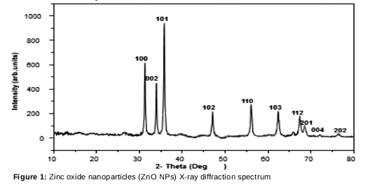
XRD spectrum of ZnO NPs
XRD results showed that ZnO NPs were present in wurtzite form, in agreement with the Joint Committee Powder Diffraction Standards (JCPDS) 89-7102 illustrated in [18-20]. The average size of the crystals was 25 nm.
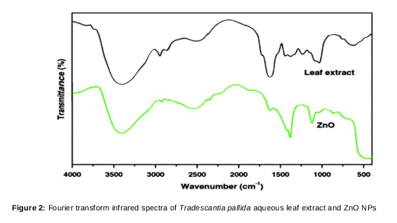
FT-IR spectrum of the ZnO NPs
The functional groups responsible for the conversion of metal precursors into metal NPs were identified using FT-IR analysis. The ZnO NPs were compared to TPALE. The peak at 1450–1500 cm−1 corresponds to N–H stretching vibrations. ZnO NP stretching was identified at 400–800 cm−1, O-H stretching at 3433 cm−1, and aldehyde C–H stretching at 2934 cm−1. A protein peak was observed at 1250 – 1270 cm−1; thus, the ZnO NPs were covered with a layer of primary and secondary metabolites, i.e., proteins and functional groups, as illustrated in [21].
Morphological and EDAX analysis
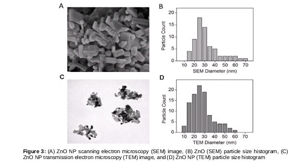
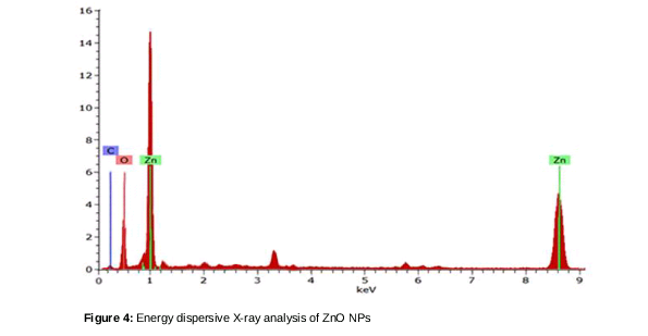
Morphological analyses were performed using SEM and TEM to identify the size and structure of the TPALE-mediated synthesized ZnO NPs. The resulting agglomerated particles were rod-shaped and had a size of 25 ± 2 nm, as illustrated in (A–D). The EDAX spectrum shows that Zn and O were present in proportions of 83 % and 17 % respectively, as shown in .
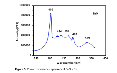
Photoluminescence of ZnO NPs
Fluorescence spectroscopy results of the ZnO NPs showed three color bands, namely, red, blue and green. Absorbance occurred at 447, 402, and 469 nm, corresponding to the blue bands, and 483 nm corresponding to the green band. The blue bands were caused by defects in the ZnO crystals, and the green band was caused by the oxygen transition vacancy, as shown in . [22,23].
Cytotoxicity activity
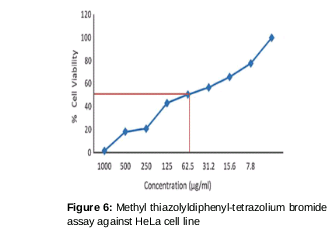
The cytotoxicity assay results showed that 98.9 % of the cancer cells died after being subjected to a high dose 1000 µg/mL of ZnO NPs. We determined the concentration required for 50 % inhibition of viability (IC50) graphically. The IC50 of ZnO NPs was 62.5 µg/mL for the HeLa cervical cancer cell line. Cells from the HeLa cell line were treated with various concentrations of ZnO NPs for 48 h. UV-visible spectroscopy was used to determine the proportion of live cells. The absorbance peak at 570 nm is clearly shown in [18].
Discussion
There has been great interest in NP synthesis based on plants, as there are several drawbacks to physical- and chemical-mediated NP synthesis. TPALE has not been used previously to synthesize NPs. Here, we synthesized ZnO NPs using TPALE and conducted cytotoxicity tests against HeLa cells. The ZnO NPs had a size of 25 nm and an agglomerated rod shape.
The present results indicate that TPALE-mediated ZnO NPs are toxic to HeLa cells, killing 98.9 % of the cancer cells when a high dose of 1000 µg/mL was used. The concentration required for IC50, determined via graphic interpolation, was 62.5 µg/mL for the HeLa cervical cancer cell line. There are several existing reports detailing the green synthesis of ZnO NPs from plant sources. These sources include Citrus aurantifolia peel, which resulted in prism-shaped particles with an average size of 35 nm [24], Vitex negundo L. extract, Caralluma fimbriata, and Euphorbia Jatropa latex [25,26]; however, few reports detail cytotoxicity assay and photoluminescence investigations of green-synthesized ZnO NPs. In future studies, it is intended that a simpler method to synthesize ZnO NPs will be developed. Such a method would serve as a reference for researchers to develop other methods to ecologically synthesize metal NPs for potential application as treatments for various medical disorders.
Conclusion
A simple approach for ZnO NP synthesis based on TPALE has been developed successfully. The mean particle size of the ZnO NPs was 25 nm, and the final product consisted of agglomerated rod-shaped particles. The synthesized nanoparticles demonstrate good luminescence properties. Cytotoxicity results against cervical cancer lines indicate 98.9 % of cancer cells died after exposure to a dose of 1000 µg/mL ZnO NPs.
Declarations
Acknowledgement
References
Archives


News Updates